
Bryozoa are aquatic colonial animals, which are abundant in modern marine environments, and have been important components of the fossil record. In places, the skeletal remains are so abundant that the fossils become an important rock-forming material. If you need a common name, then you can call them 'sea mats', 'moss animals' or 'lace corals' for some forms. The majority are marine, although brackish-water and freshwater forms are moderately common.
The word 'colonial' needs more explanation. It is applied to types of animals which reproduce (among other ways) by budding new parts asexually from the original animal. These new additions contain functioning individuals, capable of feeding independently, yet remaining attached to the rest of the the animal. Colonial animals include corals and hydroids, ascidians, graptolites and pterobranchs as well as bryozoans. Additionally, there is debate about coloniality in sponges. Bryozoan colonies have a superficial similarity with corals, but the anatomy of the bryozoan animal is much more complex.
The entire complex is called a 'colony'; the individual functional units can be called 'zooids', although 'polyp' is used for coral individuals, and 'theca' in the case of graptolites.
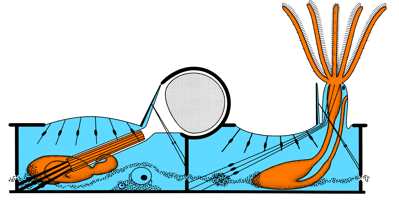
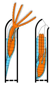
This link is a diagram of the structures of the zooids of simple bryozoans. An excellent photo of the ring of tentacles, or lophophores, of three zooids is at the Alien Life Forms page at the Connecticut River Home Page.
The earliest bryozoans are from the Early Ordovician
Go to an account of the characters of the main classes and orders of Bryozoa.
Another explanation page at the International Bryozoology Association site at London.
'Was Sind Bryozoa?'- an introduction with numerous colour photos at the Senckenberg Museum, Frankfurt
Introduction to the Bryozoans - at the Smithsonian Marine Station at Fort Pierce, Florida.